High Resolution Imaging Through Ultra Thin Optical Fibers
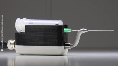
Imaging at the tip of a needle.
Scientists have developed a new technique that could revolutionize medical imaging procedures using ultra thin optical fiber and light.
Researchers at the University of Exeter in England have developed a technique to image tissues through an ultra thin optical fiber, potentially allowing for high-resolution imaging of single cells within the body.
These ultra-thin fibers hold much promise for the next generation of medical endoscopes – enabling high-resolution imaging deep inside the body at the tip of a needle.
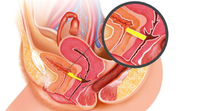
If developed further, the technique could help clinicians identify diseased cells within the body, and help with the accurate placement of needles to obtain biopsy samples.
Endoscopy is an invaluable technique that allows clinicians to peer inside our bodies while causing minimal damage. Conventional endoscopes are millimeters wide and have limited resolution – so cannot be used to inspect individual cells.
Single optical fibers are approximately 10x narrower and can enable much higher-resolution imaging – enough to examine the features of individual cells directly inside living tissue. It is normally only possible to view cells once they have been taken outside the body and placed in a microscope.
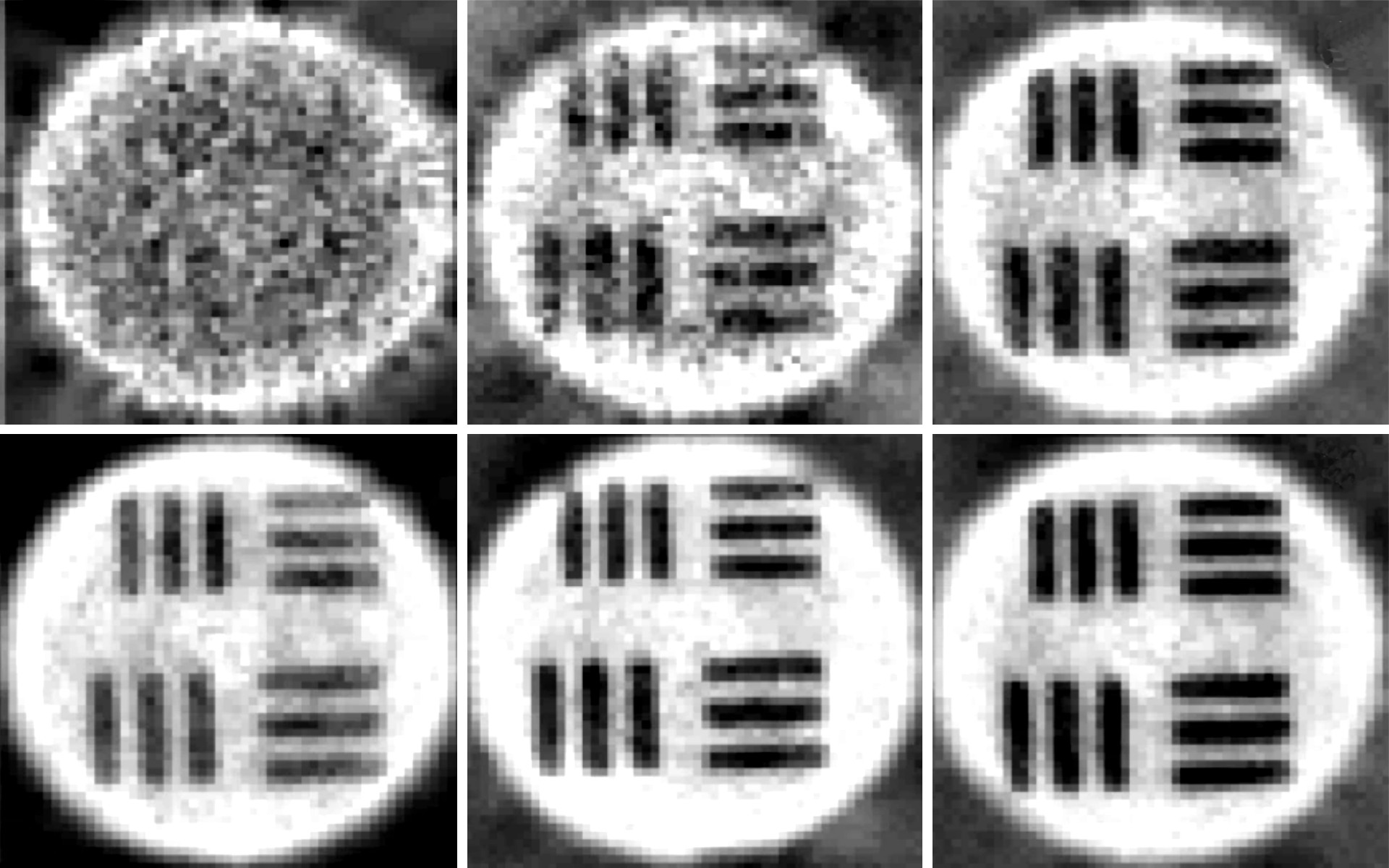
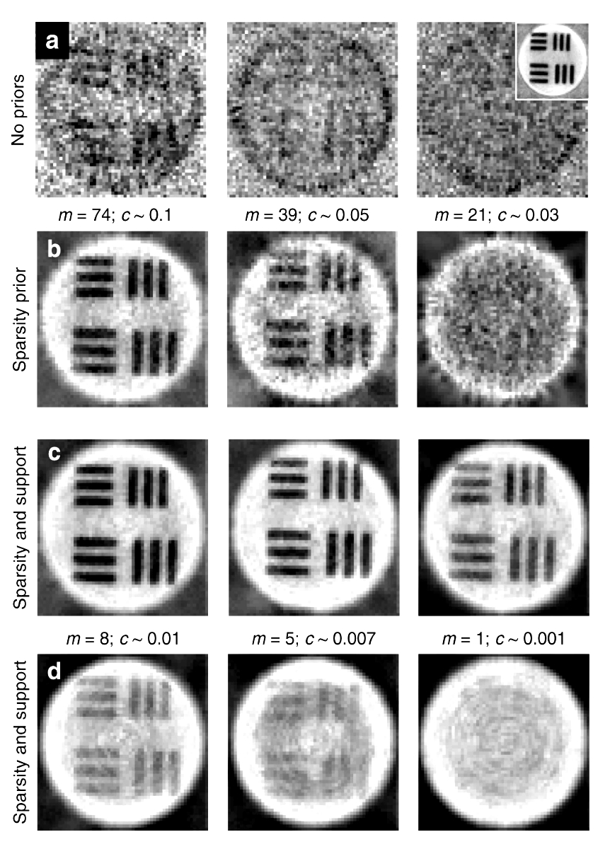
The new technique employs tiny optical fibers, the size of a human hair. The catch is that we can’t directly look through optical fibers, as they scramble the light sent through them.
This problem can be solved by unscrambling the signal to form a legible image by first understanding how the fiber distorts the light and then working backward to identify specific calibration information that can decipher the image. The researchers refer to this calibration information as a ‘key’ and recently developed a way to calculate it very quickly.
Earlier this year, Dr. Phillips’ group developed a way to measure this key extremely rapidly, in collaboration with researchers from Boston University in the USA, and the Liebniz Institute of Photonic Technologies in Germany.
The image distortion also changes depending on how the flexible fiber is bent or twisted, meaning that insertion into the body would be unlikely to render a decent image since the fiber is bound to flex during this process. To overcome this problem, the Exeter-based team has now developed a new way to keep track of how the image unscrambling key changes while the fiber is in use.
This provides a way to maintain high resolution imaging even as a single fiber-based micro-endoscope flexes. The researchers achieved this by borrowing a concept used in astronomy to see through atmospheric turbulence and applying it to look through optical fibers.
The method relies on a ‘guide-star’ – which in their case is a small brightly fluorescing particle on the end of the fiber. Light from the guide-star encodes how the key changes when the fiber bends, thus ensuring imaging is not disrupted.
This is a key advance for the development of flexible ultra-thin endoscopes. Such imaging devices could be used to guide biopsy needles to the right place, and help identify diseased cells within the body.
Dr. Phillips, an Associate Professor in the Physics and Astronomy department at the University of Exeter, said: “We hope that our work brings the visualization of sub-cellular processes deep inside the body a step closer to reality – and helps to translate this technology from the lab to the clinic.”
The latest work is in collaboration with researchers at the Liebniz Institute of Photonic Technologies in Germany, and the Brno Institute of Scientific Instruments in the Czech Republic. The project was made possible with funding from the Royal Academy of Engineering and the Royal Society in the UK, the European Research Council, and the Chinese Scholarship Council.
Primary Reference:
- Memory effect assisted imaging through multimode optical fibres. Shuhui Li, Simon A. R. Horsley, Tomáš Tyc, Tomáš Čižmár, David B. Phillips. Nature Communications volume 12, Article number: 3751 (2021)