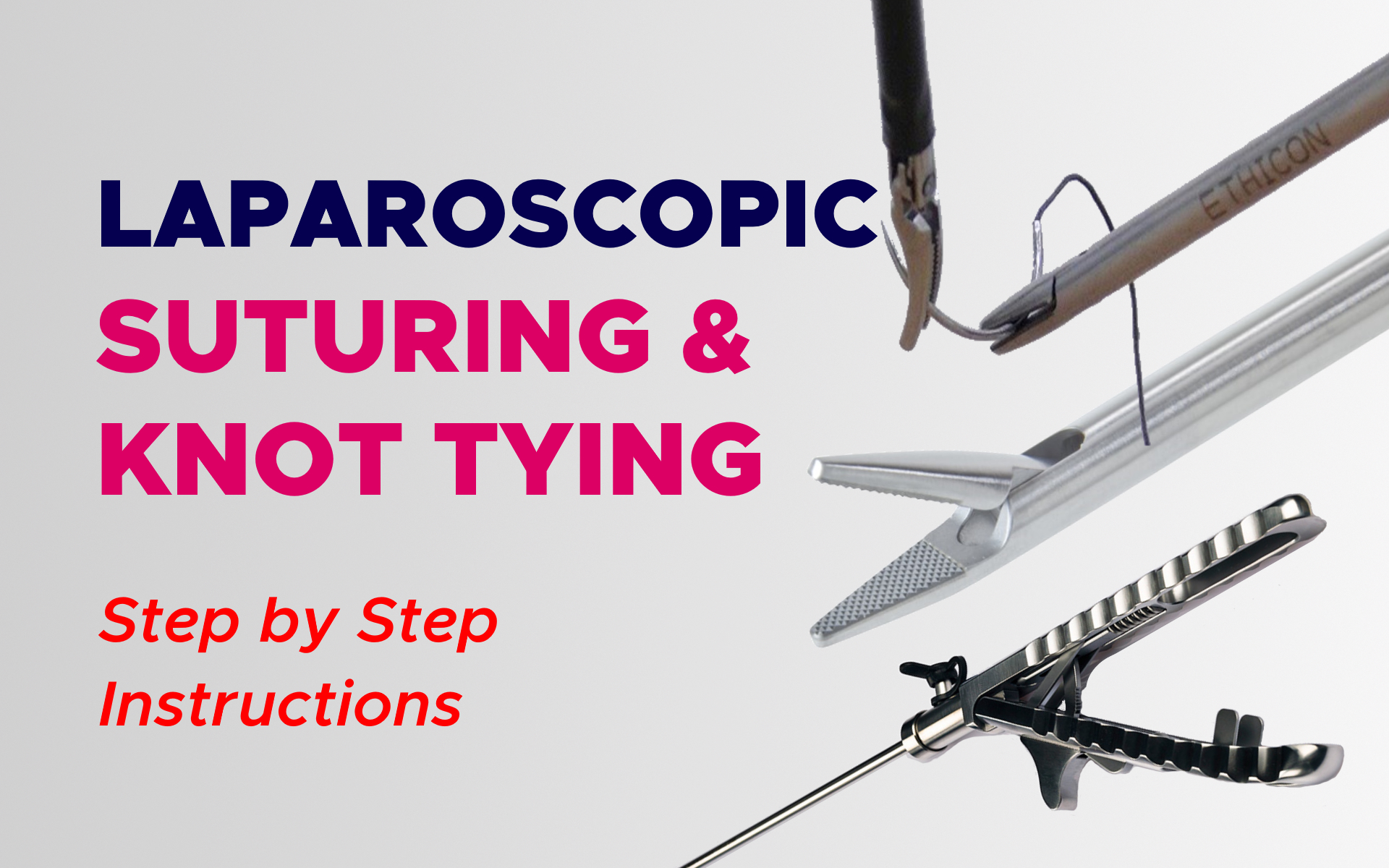
Fundamentals Of Laparoscopic Suturing And Knot Tying
Step by Step Instructions for Laparoscopic Suturing
Laparoscopic suturing and knot tying is a challenging skill to learn, but once mastered, most other laparoscopic tasks come with ease. Complex laparoscopic procedures are technically demanding with an extended learning curve.
Although tissue approximation can be achieved by various means, laparoscopic suturing remains indispensable for the execution of surgical operations.
However, suturing techniques learned in open surgery are not automatically transferred into laparoscopic suturing skills due to the visual constraints and mechanical difficulties and it requires different techniques, skills, and practice to master.
Needle Anatomy
Suture needles have several mechanical parameters for surgeons to consider. The needle point, body, and swage are important landmarks of the needle. The swage is the end of the needle that is connected to the suture. The chord length is defined as the direct distance from the needle’s point to the swage.
This measurement should not be confused with length, which is the distance along the curvature of the needle from the swage to the point.
The extent of curvature for the needle is defined as the fraction of a circle that the needle measures (eg, 1/4 circle or 5/8 circle). The radius is measured as the radius of the circle by which the extent of curvature is defined.

Needle Entrance (Proper Loading and Back Loading)
Laparoscopic introduction of curved needles into the peritoneal cavity maybe a slightly more involved affair. lf 10 mm Operating trocars are used, then these curved needles may be inserted and later removed easily.
However, if only small trocars (5mm) are available during surgery, then these curved needles must be inserted using a different technique.
These curved needles must first be preloaded in the 55 mm trocars before all 3 (trocar, needle holder, and needle) are introduced into the peritoneal cavity. First, the needle holder is passed through the trocar and this is then used to grasp the tail end of the suture.
The needle is then ‘back-loaded, by pulling the suture through the trocar sleeve. Next, the tail .end of the suture is then released, and the needle holder reintroduced into the trocar.
The suturing device is then used to regrasp the suture 1-2 cm from the swage point. The loaded needle holder trocar complex is now ready to be introduced into the peritoneal cavity.
Loading the Needle Within the Needle Driver
There are two processes for loading the needle. The option depends upon the precise conditions and also the proximity or otherwise of a smooth serosal surface:
- The deposit – pick-up technique
- The dangling pirouette technique
If adjustment of the needle is needed, this can be done in 3 ways:
Maintaining a light grip about the needle and pulling the thread taut so that angle from the needle is modified
Maintaining an easy grip and brushing the end from the needle gently from the close by tissues forward for backward within the 3 o’clock direction
Reducing the grip from the needle and gently hooking it on the superficial serosal layer in an avascular area. A popular but inefficient technique involves while using supporting the assisting instrument to reposition the angle from the needle.
Throwing a Stitch, Needle Driving Principles
Angle of approach and direction of the force: The end of the needle must approach tissue at right angles regardless of the configuration of the needle. The direction from the power apply with the needle driver should be perpendicular to the cut surface or tissue edge.
Needle detection: Needle detection occurs when the needle is not pushed directly opposite to the plane of tissue resistance and the extent of detection is direct of the power in the perpendicular interface with the resistance provided by the tissue.
Counter pressure in needle driving: The correct using counter pressure or counter traction expedites suturing.
Technique of needle driving: The needle driver may be the dominant-handed instrument and the assistant grasper is grasped.
Needle positioning: A slowly driven needle passage results in a smoother and much more successful passage. The needle is driven having a delicate grip and also the direction from the pushing force is maintained directly against tissue resistance.
Entrance bite: for that entrance bite, the needle tip should be ready that’s perpendicular, or nearly so, towards the tissue surface. The motive force will be supinated by rotating the needle clockwise, thereby pushing it down with the tissues and then forward and upwards penetrating appropriate tissue layers.
Extracting the needle: two methods are usually used one, once the needle tip is grasped by the assisting instrument when only the tip from the needle is shown. Second, whenever a half circle needle is used, it’s just extracted after extensive rotation and not along the horizontal plane.
Exit bite: the needle ought to be pulled from the tissue surface only for ten or twenty yards enough to make it possible for the maneuverability required for reloading the needle within the driver.
Knot Tying
An ideal stitch is a which supports the tissue edges as well as correct tension and resists reverse slippage.
The three procedures in knotting:
- Tying the knot (configuration)
- Working or drawing the knot (shaping)
- Snuggling or locking the knot (securing)
All of the three stages are essential along with a knot is secure only if it is tied correctly, drawn (shaped) to adapt to the anatomy of the knot, and locked tightly. As Clifford Ashely wrote ‘a knot is never nearly right; it is either exactly right or is hopelessly wrong’.
The perfect characteristics of the surgical knot are:
- It ought to be safe
Triadic relationship of port sites. In suturing placed in Endoscopic surgery it is advisable to always maintain some relationship between your both sides. The telescope and suturing port sites form a triad with the optical port within the center and suturing ports on each side.
Wrapping processes for knotting: There are two basic techniques of wrapping most commonly practiced by Endoscopic surgeons.
- Overwrap method
- Underwrap method
Essentials in Intracorporeal Knot Tying
A period of ligature materials for trying a knot (just like a rope) can be considered as consisting of three sections:
- the end
- the bight
- the standing part
The essentials that ought to be followed while intracorporeal knotting is listed below:
– Good magnification
– Economy of motion
– Directional hold principle
– Avoidance of instrument crossing
– Correct knot-tying choreography
– Execution of the knots near the tissue surfaces
– Closed instrument jaws except during grasping
– Awareness of dominant versus assisting instruments throughout the knotting process
– Correct wrapping techniques
– Knots configured and tied near to the tail
Extracorporeal Knots
External or extracorporeal slip knots are used in endoscopic surgery for:
– Ligature of vessels and tubular structures in continuity
– Transfixation of huge vascular pedicles
– Interrupted suturing with external knots
In all these situations, the knot is tied and drawn outside the body after which it ‘slipped down’ with a knot pusher towards the intended target after which stiffened by traction on the standing up part against the knot pusher.
Extracorporeal versus Intracorporeal knots:
More often than not during endoscopic surgery, intracorporeal knotting utilizing instrument-tied knots is preferred to extracorporeal tying. However, in the next situations, extracorporeal knotting is recommended:
- Ligature in the continuity of large vessels
- Suturing in regions of limited access where the performing space is restricted
- In the approximation of edges of defects where the force necessitates approximating the edges is substantial
Rules Governing External Slip Knotting
The following rules should be followed for a safe ligature for slip knots in endoscopic surgery are:
– Type of the thread must be 1.5 m and also the gauge ought to be 2/0 or greater.
– The type of slip knot selected depends upon the ligature material being used. Certain slip knots provide sufficient holding strength with catgut but not along with other materials.
– For any ligature material, the holding force (resistance to reverse slipping) of any surgical slip knot varies directly using its caliber. Thus the holding strength of a 1/0 slip knot is roughly twice those of the 2/0 equivalent.
– Stiff hydrophobic monofilament material ought to be avoided since it exerts a lesser frictional hold and has a larger tendency to spill than braided.

Hi, can you please add me to your Laparoscopy Course. My email seen at the belle, thanks
Хотел бы у Вас узнать, какие камеры Вы используете на тренажёрах?