Uterine Cancer: Introduction
Definition, Anatomy, Precancerous Lesions, Hyperplasia and Types of Uterine Cancer
Uterine cancer is the most common cancer occurring within a woman’s reproductive system. Uterine cancer begins when healthy cells in the uterus change and grow out of control, forming a mass called a tumor. More than 90% of uterine cancers occur in the endometrium. The majority of women with uterine cancer are diagnosed at an early stage, largely due to the presence of abnormal vaginal bleeding as an early symptom.
Uterus
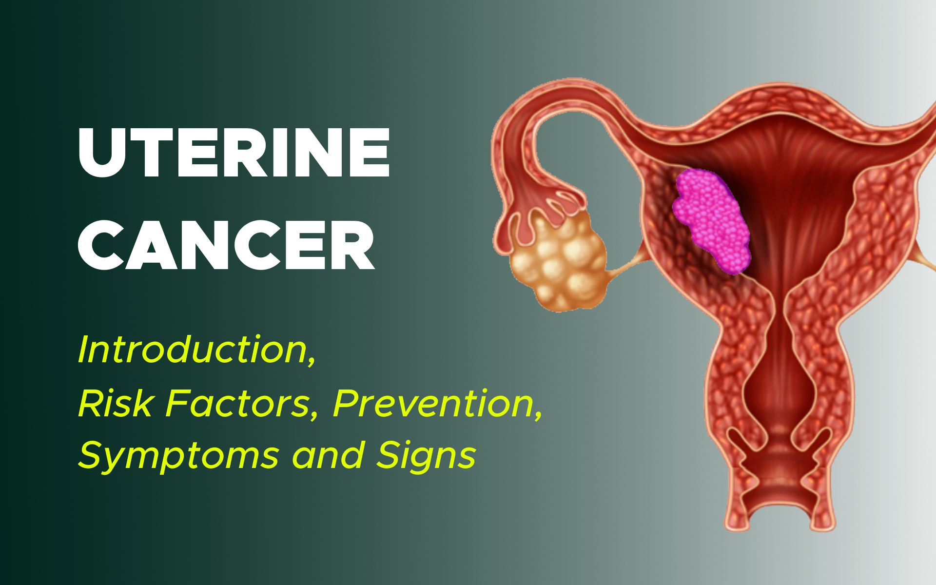
The pear-shaped uterus is hollow and located in a woman’s pelvis between the bladder and rectum. The uterus is also called the womb. It is where a baby grows when a woman is pregnant. The uterus has 3 sections:
The cervix, which is the narrow lower section
The isthmus, which is the broad section in the middle
The fundus, which is the dome-shaped top section.
The uterus is made up of 3 layers: the endometrium (inner layer), the myometrium (the thickest layer composed almost entirely of muscle), and the serosa (the thin outer lining of the uterus).
During a woman’s childbearing years, her ovaries typically release an egg every month, and the endometrium grows and thickens in preparation for pregnancy. If the woman does not get pregnant, this endometrial lining passes out of her body through her vagina, a process known as menstruation. This process continues until menopause when a woman’s ovaries stop releasing eggs.
Uterine Cancer
A tumor can be cancerous or benign. A cancerous tumor is malignant, meaning it can grow and spread to other parts of the body. A benign tumor can grow but generally will not spread to other body parts.
Noncancerous conditions of the uterus include:
Fibroids: Benign tumors in the muscle of the uterus
Benign polyps: Abnormal growths in the lining of the uterus
Endometriosis: A condition in which endometrial tissue, which usually lines the inside of the uterus, is found on the outside of the uterus or other organs
Endometrial hyperplasia: A condition in which there is an increased number of cells and glandular structures in the uterine lining. Endometrial hyperplasia can have either normal or atypical cells and simple or complex glandular structures. The risk for developing cancer in the lining of the uterus is higher when endometrial hyperplasia has atypical cells and complex glands.
There are 2 major types of uterine cancer:
Adenocarcinoma:
This type makes up more than 80% of uterine cancers. It develops from cells in the endometrium. This cancer is commonly called endometrial cancer. One common endometrial adenocarcinoma subtype is called endometrioid carcinoma.
Treatment for this type of cancer varies depending on the grade of the tumor, how far it goes into the uterus and the stage or extent of the disease. Less common subtypes of uterine adenocarcinomas include serous, clear cell, and carcinosarcoma. Carcinosarcoma is a mixture of adenocarcinoma and sarcoma (see below).
Sarcoma:
This type of uterine cancer develops in the supporting tissues of the uterine glands or in the myometrium, which is the uterine muscle. Sarcoma accounts for about 2% to 4% of uterine cancers. Subtypes of endometrial sarcoma include leiomyosarcoma, endometrial stromal sarcoma, and undifferentiated sarcoma.
Cancer confined to the uterine cervix is treated differently from uterine cancer. The rest of this section covers the more common endometrial (adenocarcinoma) cancer.
