Thyroid Cancer
Thyroid Cancer: Introduction, Risk Factors, Symptoms, Signs and Diagnosis
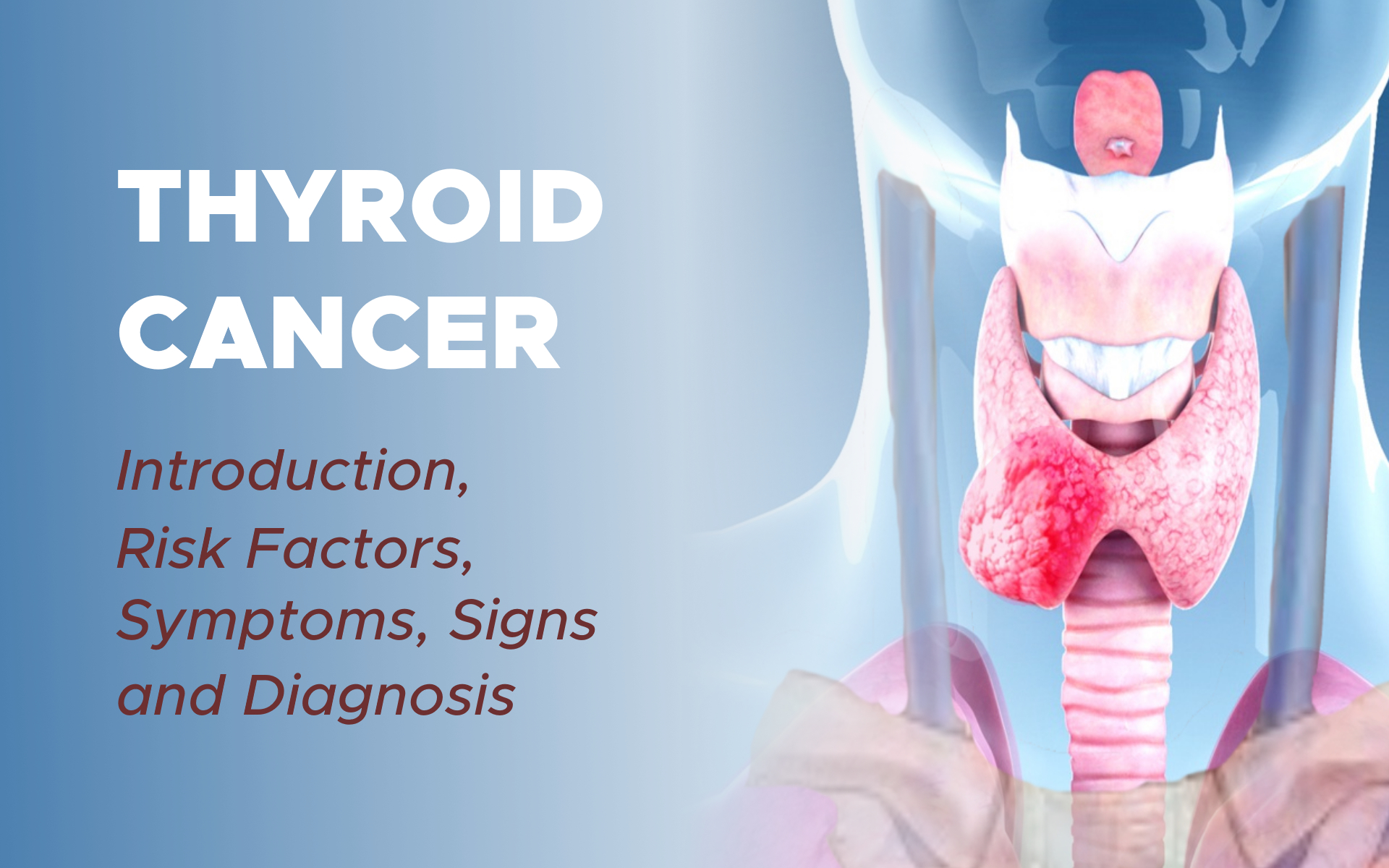
Thyroid cancer begins in the thyroid gland. Thyroid gland is located in the front of the neck just below the larynx, which is called the voice box. The thyroid gland is part of the endocrine system, which regulates hormones in the body. The thyroid gland absorbs iodine from the bloodstream to produce thyroid hormones, which regulate a person’s metabolism.
A normal thyroid gland has 2 lobes, 1 on each side of the windpipe, joined by a narrow strip of tissue called the isthmus. A healthy thyroid gland is barely palpable, which means it is hard to find by touch. If a tumor develops in the thyroid, it is felt as a lump in the neck.
A swollen or enlarged thyroid gland is called a goiter, which may be caused when a person does not get enough iodine. However, most Americans receive enough iodine from salt, and a goiter under these circumstances is caused by other reasons.
Thyroid Tumors
Thyroid cancer starts when healthy cells in the thyroid change and grow out of control, forming a mass called a tumor. The thyroid gland contains 2 types of cells:
1. Follicular Cells
These cells are responsible for the production of thyroid hormone. Thyroid hormone is needed to live. The hormone controls the basic metabolism of the body. It controls how quickly calories are burned.
This can affect weight loss and weight gain, slow down or speed up the heartbeat, raise or lower body temperature, influence how quickly food moves through the digestive tract, control the way muscles contract, and control how quickly dying cells are replaced.
2. C Cells
These special cells of the thyroid make calcitonin, a hormone that participates in calcium metabolism.
A tumor can be cancerous or benign. A cancerous tumor is malignant, meaning it can grow and spread to other parts of the body. A benign tumor means the tumor can grow but will not spread.
Thyroid tumors can also be called nodules, and about 90% of all thyroid nodules are benign.
Types of Thyroid Cancer
There are 5 main types of thyroid cancer:
1. Papillary Thyroid Cancer

Papillary thyroid cancer develops from follicular cells and usually grow slowly. It is the most common type of thyroid cancer. It is usually found in 1 lobe. Only 10% to 20% of papillary thyroid cancer appears in both lobes.
It is a differentiated thyroid cancer, meaning that the tumor looks similar to normal thyroid tissue under a microscope. Papillary thyroid cancer can often spread to lymph nodes.
2. Follicular Thyroid Cancer
Follicular thyroid cancer also develops from follicular cells and usually grows slowly. Follicular thyroid cancer is also a differentiated thyroid cancer, but it is far less common than papillary thyroid cancer. Follicular thyroid cancer rarely spreads to lymph nodes.
Follicular thyroid cancer and papillary thyroid cancer are the most common differentiated thyroid cancers. They are very often curable, especially when found early and in people younger than 50. Together, follicular and papillary thyroid cancers make up about 95% of all thyroid cancer.
3. Hurthle Cell Cancer
Hurthle cell cancer, also called Hurthle cell carcinoma, is cancer that is arises from a certain type of follicular cell. Hurthle cell cancers are much more likely to spread to lymph nodes than other follicular thyroid cancers.
4. Medullary Thyroid Cancer (MTC)
MTC develops in the C cells and is sometimes the result of a genetic syndrome called multiple endocrine neoplasia type 2 (MEN2). This tumor has very little, if any, similarity to normal thyroid tissue. MTC can often be controlled if it is diagnosed and treated before it spreads to other parts of the body.
MTC accounts for about 3% of all thyroid cancers. About 25% of all MTC is familial. This means that family members of the patient will have a possibility of a similar diagnosis. The RETproto-oncogene test can confirm if family members also have familial MTC (FMTC).
5. Anaplastic Thyroid Cancer
This type is rare, accounting for about 1% of thyroid cancer. It is a fast-growing, poorly differentiated thyroid cancer that may start from differentiated thyroid cancer or a benign thyroid tumor.
Anaplastic thyroid cancer can be subtyped into giant cell classifications. Because this type of thyroid cancer grows so quickly, it is more difficult to treat successfully.
In addition, other types of cancer may start in or around the thyroid gland.
Risk Factors
A risk factor is anything that increases a person’s chance of developing cancer. Although risk factors often influence the development of cancer, most do not directly cause cancer.
Some people with several risk factors never develop cancer, while others with no known risk factors do. Knowing your risk factors and talking about them with your doctor may help you make more informed lifestyle and health care choices.
The following factors may raise a person’s risk of developing thyroid cancer:
1. Gender
Women are diagnosed with 3 of every 4 thyroid cancers.
2. Age
Thyroid cancer can occur at any age, but about two-thirds of all cases are found in people between the ages of 20 and 55. Anaplastic thyroid cancer is usually diagnosed after age 60. Older infants (10 months and older) and adolescents can develop MTC, especially if they carry the RET proto-oncogene mutation (see below).
3. Genetics
Some types of thyroid cancer are associated with genetics. Below are some key facts about this disease, genes, and family history.
An abnormal RET oncogene, which can be passed from parent to child, may cause MTC. Not everyone with an altered RET oncogene will develop cancer. Blood tests and genetic tests can detect the gene.
Once the altered RET oncogene is identified, a doctor may recommend surgery to remove the thyroid gland before cancer develops.

People with MTC are encouraged to have genetic testing to determine if a mutation of the RET proto-oncogene is present. If so, genetic testing of parents, siblings, and children will be recommended.
- A family history of MTC increases a person’s risk. People with MEN2 syndromeare also at risk for developing other types of cancers.
- A family history of precancerous polyps in the colon, also called the large intestines, increases the risk of developing papillary thyroid cancer.
4. Radiation Exposure
Exposure to moderate levels of radiation to the head and neck may increase the risk of papillary and follicular thyroid cancers. Such sources of exposure include:
- Low-dose to moderate-dose x-ray treatments used before 1950 to treat children with acne, tonsillitis, and other head and neck problems.
- Radiation therapy for Hodgkin lymphoma or other forms of lymphoma in the head and neck.
- Exposure to radioactive iodine, also called I-131 or RAI, especially in childhood.
- Exposure to ionizing radiation, including radioactive fallout from atomic weapons testing during the 1950s and 1960s and nuclear power plant fallout. Examples include the 1986 Chernobyl nuclear power plant accident and the 2011 earthquake that damaged nuclear power plants in Fukushima, Japan. Another source of I-131 is environmental releases from atomic weapon production plants.
5. Diet Low in Iodine
Iodine is needed for normal thyroid function. In the United States, iodine is added to salt to help prevent thyroid problems.
6. Race
White people and Asian people are more likely to develop thyroid cancer, but this disease can affect a person of any race or ethnicity.
7. Breast Cancer
A recent study showed that breast cancer survivors may have a higher risk of thyroid cancer, particularly in the first 5 years after diagnosis and for those diagnosed with breast cancer at a younger age. This finding continues to be examined by researchers.
Symptoms and Signs
It is common for people with thyroid cancer to have few or no symptoms. Thyroid cancers are often diagnosed by routine examination of the neck during a general physical exam.
They are also unintentionally found by X-Rays or other imaging scans that were performed for other reasons. People with thyroid cancer may experience the following symptoms or signs.
Sometimes, people with thyroid cancer do not have any of these changes. Or, the cause of a symptom may be a different medical condition that is not cancer.
- A lump in the front of the neck, near the Adam’s apple
- Hoarseness
- Swollen glands in the neck
- Difficulty swallowing
- Difficulty breathing
- Pain in the throat or neck
- A cough that persists and is not caused by a cold
If you are concerned about any changes you experience, please talk with your doctor. Your doctor will ask how long and how often you’ve been experiencing the symptom(s), in addition to other questions. This is to help figure out the cause of the problem, called a diagnosis.
These symptoms may be caused by thyroid cancer; other thyroid problems, such as a goiter; or a condition not related to the thyroid, such as an infection.
If cancer is diagnosed, relieving symptoms remains an important part of cancer care and treatment. This may be called palliative care or supportive care.
It is often started soon after diagnosis and continued throughout treatment. Be sure to talk with your health care team about the symptoms you experience, including any new symptoms or a change in symptoms.
Diagnosis
Doctors use many tests to find, or diagnose, cancer. They also do tests to learn if cancer has spread to another part of the body from where it started. If this happens, it is called metastasis.
For example, imaging tests can show if the cancer has spread. Imaging tests show pictures of the inside of the body. Doctors may also do tests to learn which treatments could work best.
For most types of cancer, a biopsy is the only sure way for the doctor to know if an area of the body has cancer. In a biopsy, the doctor takes a small sample of tissue for testing in a laboratory. If a biopsy is not possible, the doctor may suggest other tests that will help make a diagnosis.
Your doctor may consider these factors when choosing a diagnostic test:
- The type of cancer suspected
- Your signs and symptoms
- Your age and general health
- The results of earlier medical tests
This section describes options for diagnosing thyroid cancer. Not all tests listed below will be used for every person.
1. Physical Examination
The doctor will feel the neck, thyroid gland, throat, and lymph nodes (the tiny, bean-shaped organs that help fight infection) in the neck for unusual growths or swelling. If surgery is recommended, the larynx may be examined at the same time with a laryngoscope, which is a thin, flexible tube with a light.
2. Blood Tests
There are several types of blood tests that may be done during diagnosis and to monitor the patient during and after treatment. This includes tests called tumor marker tests. Tumor markers are substances found at higher-than-normal levels in the blood, urine, or body tissues of some people with cancer.
a. Thyroid Hormone Levels
As explained in the Introduction, thyroid hormones regulate a person’s metabolism. The doctor will use this test to find out the current levels of the thyroid hormones triiodothyronine (T3) and thyroxine (T4) in the body.
b. Thyroid-Stimulating Hormone (TSH)
This blood test measures the level of TSH, a hormone produced by the pituitary gland near the brain. If the body is in need of thyroid hormone, the pituitary gland releases TSH to stimulate production.
c. Tg and TgAb
Thyroglobulin (Tg) is a protein made naturally by the thyroid as well as by differentiated thyroid cancer. After treatment, there should be very low levels of thyroglobulin in the blood since the goal of treatment is to remove all thyroid cells.
If Tg is rising after surgery and/or radioactive iodine, it may be a sign of more cancer. A tumor marker test may be done to measure the body’s Tg level before, during, and/or after treatment.
There is also a test for thyroglobulin antibodies (TgAb), which are proteins produced by the body to attack thyroglobulin that occur in some patients. If TgAb is found, it is known to interfere with the results of the Tg level test.
d. Medullary Type-Specific Tests
If MTC is a possibility, the doctor will order tumor marker tests to check for high calcitonin and carcinoembryonic antigen (CEA) levels. The doctor should also recommend a blood test to detect the presence of RETproto-oncogenes, particularly if there is a family history of MTC.
3. Ultrasound
An ultrasound uses sound waves to create a picture of the internal organs. An ultrasound wand or probe is guided over the skin of the neck area.
High-frequency sound waves create a pattern of echoes that show the doctor the size of the thyroid gland and specific information about any nodules, including whether a nodule is solid or a fluid-filled sac called a cyst.
4. Molecular Testing of the Nodule Sample
Your doctor may recommend running laboratory tests on a tumor sample to identify specific genes, proteins, and other factors unique to the tumor.
Genetic analysis of your thyroid nodule may allow you to understand the risk of the thyroid nodule being cancerous. Other genetic, protein, and molecular analysis of thyroid cancers can help determine your treatment options, including types of treatments called targeted therapy.
5. Radionuclide Scanning
This test may also be called a whole-body scan. This scan will either be done using a very small, harmless amount of radioactive iodine I-131 or I-123, called a tracer. It is used most often to learn more about a thyroid nodule. In this test, the patient swallows the tracer, which is absorbed by thyroid cells.
This makes the thyroid cells appear on the scan image, allowing the doctor to see differences between those cells and other body structures.
6. X-Ray
An x-ray is a way to create a picture of the structures inside of the body, using a small amount of radiation. For instance, a chest x-ray can help doctors determine if the cancer has spread to the lungs.
7. Computed Tomography (CT or CAT) Scan
A CT scan creates a 3-dimensional picture of the inside of the body using x-rays taken from different angles. A computer combines these pictures into a detailed, cross-sectional view that shows any abnormalities or tumors.
A CT scan can be used to measure the tumor’s size. Sometimes, a special dye called a contrast medium is given before the scan to provide better detail on the image. This dye can be injected into a patient’s vein or given as a pill to swallow.
CT scans are often used in people with thyroid cancer to examine parts of the neck that cannot be seen with ultrasound (see above). Also, CT scans of the chest may be needed to look to see if thyroid cancer has spread to that area of the body.
CT scans of the abdomen may be used to see if thyroid cancer has spread to the liver or other sites. Patients with the hereditary form of medullary thyroid cancers may be at risk for developing other types of endocrine tumors in the abdomen; these patients may also have a CT scan of the abdomen.
8. Positron Emission Tomography (PET) or PET-CT Scan

A PET scan is usually combined with a CT scan (see above), called a PET-CT scan. But you may hear your doctor refer to this procedure just as a PET scan. A PET scan is a way to create pictures of organs and tissues inside the body.
A small amount of a radioactive sugar substance is injected into the patient’s body. This sugar substance is taken up by cells that use the most energy. Because cancer tends to use energy actively, it absorbs more of the radioactive substance. A scanner then detects this substance to produce images of the inside of the body.
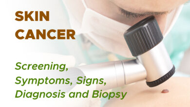
9. Biopsy
A biopsy is the removal of a small amount of tissue for examination under a microscope. Other tests can suggest that cancer is present, but only a biopsy can make a definite diagnosis. The way to determine whether a nodule is cancerous or benign is through a biopsy.
During this procedure, the doctor removes cells from the nodule that are then examined by a cytopathologist. A cytopathologist is a doctor who specializes in analyzing cells and tissue to diagnose disease. This test is often done with the help of ultrasound.
A biopsy for thyroid nodules will be done in 1 of 2 ways:
a. Fine Needle Aspiration
This procedure is usually performed in a doctor’s office or clinic. It is an important diagnostic step to find out if a thyroid nodule is benign or cancerous. A local anesthetic may be injected into the skin to numb the area before the biopsy.
The doctor inserts a thin needle into the nodule and removes cells and some fluid. The procedure may be repeated 2 or 3 times to get samples from different areas of the nodule. A report of the results of this test is created by the cytopathologist.
A pathologist is a doctor who specializes in interpreting laboratory tests and evaluating cells, tissues, and organs to diagnose disease. The test can be positive, meaning there are cancerous cells, or negative, meaning there are no cancerous cells. The test can also be undetermined, meaning it is not clear whether cancer is there.
b. Surgical Biopsy
If the needle aspiration biopsy is not clear, the doctor may suggest a biopsy in which the nodule and possibly the affected lobe of the thyroid will be removed using surgery.
Removal of the nodule alone is usually not recommended due to the potential to incompletely remove the potential malignancy without adequate margins, which is an area of tissue around the nodule.
This procedure is usually done under general anesthesia. It may also require a hospital stay.