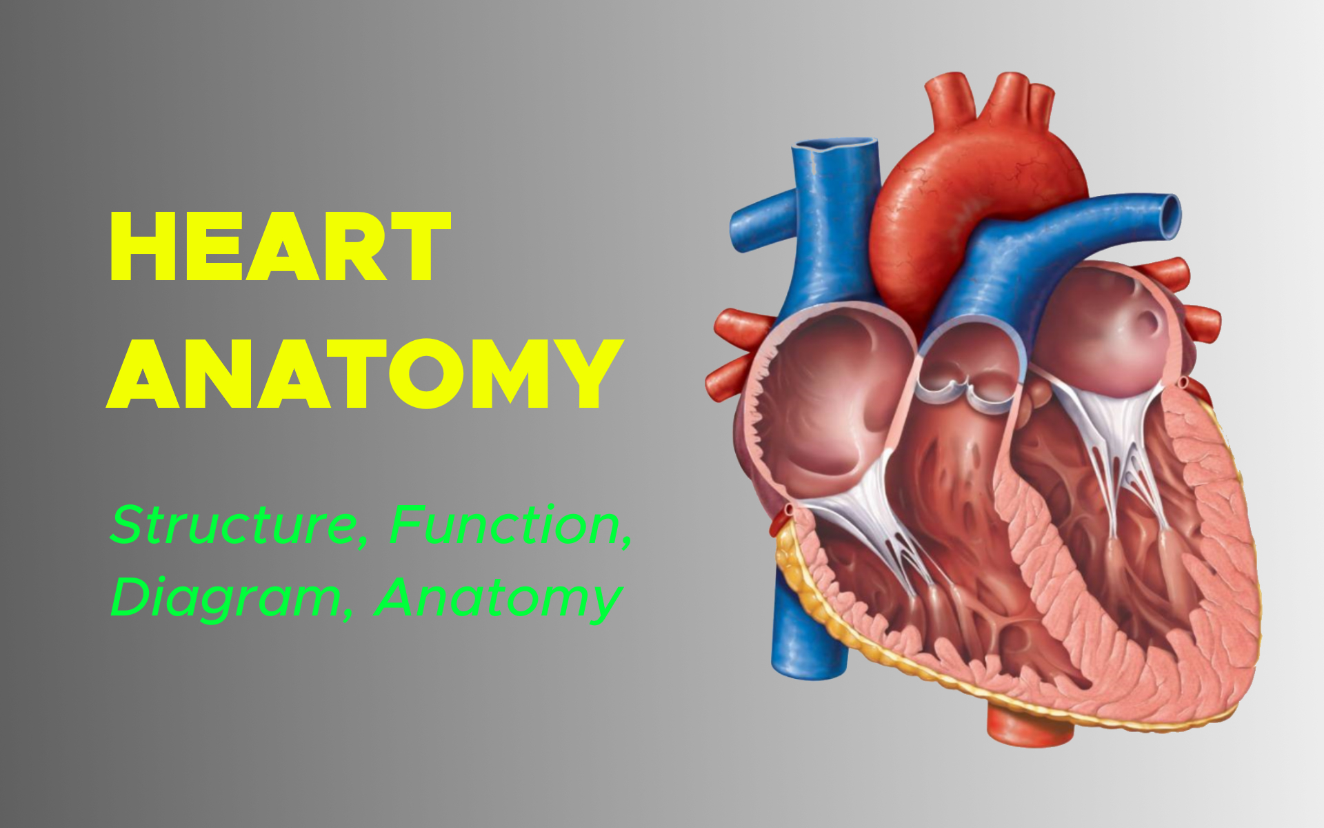
Anatomy of the Human Heart
Beats about 100,000 times, pumping 2,000 gallons of blood: That’s a lot of work for an organ no bigger than a large fist and weighing 8 to 12 ounces.
The heart is a muscular pump that provides the force necessary to circulate the blood to all the tissues in the body. Each day, the average human heart beats about 100,000 times, pumping 2,000 gallons of blood through the body. In fact, the heart does more physical work than any other muscle over a lifetime.
Its function is vital because, to survive, the tissues need a continuous supply of oxygen and nutrients, and metabolic waste products have to be removed. Deprived of these necessities, cells soon undergo irreversible changes that lead to death.
While blood is the transport medium, the heart is the organ that keeps the blood moving through the vessels. The normal adult heart pumps about 5 liters of blood every minute throughout life. If it loses its pumping effectiveness for even a few minutes, the individual’s life is jeopardized.
Located between the lungs in the middle of the chest, the heart pumps blood through the network of arteries and veins known as the cardiovascular system. It pushes blood to the body’s organs, tissues and cells.
Blood delivers oxygen and nutrients to every cell and removes the carbon dioxide and other waste products made by those cells.
Blood is carried from the heart to the rest of the body through a complex network of arteries, arterioles and capillaries. Blood is returned to the heart through venules and veins.
Layers of the Heart Wall
Three layers of tissue form the heart wall. The outer layer of the heart wall is the epicardium, the middle layer is the myocardium, and the inner layer is the endocardium.
Chambers of the Heart
The internal cavity of the heart is divided into four chambers:
- Right atrium
- Right ventricle
- Left atrium
- Left ventricle

The two atria are thin-walled chambers that receive blood from the veins. The two ventricles are thick-walled chambers that forcefully pump blood out of the heart. Differences in thickness of the heart chamber walls are due to variations in the amount of myocardium present, which reflects the amount of force each chamber is required to generate.
The right atrium receives deoxygenated blood from systemic veins; the left atrium receives oxygenated blood from the pulmonary veins.
Valves of the Heart
Pumps need a set of valves to keep the fluid flowing in one direction and the heart is no exception. The heart has two types of valves that keep the blood flowing in the correct direction.
The valves between the atria and ventricles are called atrioventricular valves (also called cuspid valves), while those at the bases of the large vessels leaving the ventricles are called semilunar valves.
The right atrioventricular valve is the tricuspid valve. The left atrioventricular valve is the bicuspid, or mitral, valve. The valve between the right ventricle and pulmonary trunk is the pulmonary semilunar valve. The valve between the left ventricle and the aorta is the aortic semilunar valve.
When the ventricles contract, atrioventricular valves close to prevent blood from flowing back into the atria. When the ventricles relax, semilunar valves close to prevent blood from flowing back into the ventricles.
Pathway of Blood through the Heart
While it is convenient to describe the flow of blood through the right side of the heart and then through the left side, it is important to realize that both atria and ventricles contract at the same time. The heart works as two pumps, one on the right and one on the left, working simultaneously.
Right and Left Sides of the Heart
The right-hand side of the heart pumps blood needing oxygen to the lungs.
This blood goes to the lungs where oxygen is loaded up and sent back to the heart. The oxygen-rich blood reaches the left side of the heart and then pumps it to where it is needed throughout the body.
Blood that supplied the muscles and tissues with its oxygen then returns to the right side of the heart to restart the cycle.
Coronary Arteries: Blood Supply to the Myocardium
The myocardium of the heart wall is a working muscle that needs a continuous supply of oxygen and nutrients to function efficiently. For this reason, cardiac muscle has an extensive network of blood vessels to bring oxygen to the contracting cells and to remove waste products.
The right and left coronary arteries, branches of the ascending aorta, supply blood to the walls of the myocardium. After blood passes through the capillaries in the myocardium, it enters a system of cardiac (coronary) veins. Most of the cardiac veins drain into the coronary sinus, which opens into the right atrium.
If the coronary arteries are narrowed by fatty deposits in the lining of the arteries (atherosclerosis), the flow of blood to the heart muscle may be restricted. If the heart muscle doesn’t get enough blood, it doesn’t get enough oxygen to work properly – it’s called ischaemia.
Ischaemia can cause chest pain or angina, often described as a feeling of pressure or tightness in the chest. Angina pain can also be felt in the neck, shoulders, or arms. Angina is often caused by physical activity and usually improves with rest.
Superior and Inferior Vena Cavae
These are the 2 large veins that enter the right side of the heart and bring low-oxygen blood into the right atrium. The superior vena cava carries blood from the head and arms and upper body; the inferior vena cava carries blood from the trunk and legs – the lower body.
Pulmonary Veins
The right and left pulmonary veins carry the oxygen-rich blood back into the left atrium from the lungs to the heart.
Pulmonary Arteries
The pulmonary arteries of the right and left branch of the main pulmonary trunk. From the right ventricle, the blood which needs oxygen is pumped into them and taken to the lungs where it is loaded with oxygen.
Aorta
The aorta is the most extensive artery in the body. The blood filled with oxygen is pumped into the aorta by the left ventricle, round the aortic arch, and out into the upper body via the 3 main arteries that branch off the aortic arch and through the descending aorta into the thorax, trunk and lower body.